
Signalment: Ruby, a 4-year-6-month-old spayed female Pembroke Welsh Corgi
Presenting Complaint: Chronic and recurrent urinary tract infections (UTIs)
History:
Ruby was adopted at approximately 2 years of age. She was first evaluated on 3/16/2023 for excessive vulvar licking and mucous discharge. While her urinalysis (UA) was unremarkable at that time, a recessed vulva was noted. She was treated symptomatically with antihistamines, antifungals, and topical hygiene. Over the next several months, she developed recurrent UTIs, with signs including hematuria, pyuria, urinary dribbling, and stranguria. Her infections were managed episodically with antibiotics and cranberry supplementation. Despite these interventions and a switch to a urinary diet (Royal Canin SO), Ruby continued to experience recurrent infections.
By October 2024, Ruby had undergone multiple courses of antibiotics for documented E. coli infections, and despite normal physical exams and unremarkable urinalyses between episodes, persistent perivulvar irritation and grooming behaviors were noted. Her history of vulvar conformation abnormality, waxing/waning urinary symptoms, and repeated infections raised concern for underlying subclinical urinary incontinence—a known predisposing factor for recurrent UTIs in female dogs.
Diagnostic Workup:
At presentation to AVES Internal Medicine, Ruby’s physical exam revealed approximately 70% hooded vulva with mild erythema and salivary staining. Bloodwork and urinalysis were within normal limits. A urinary tract ultrasound showed no abnormalities.
Cystoscopy was performed (see image documentation below). This revealed multifocal pale pink nodules in the vestibule surrounding the urethral orifice, with no vestibulovaginal septal remnant. The urethra was normal in both length and mucosal appearance. The bladder contained similar nodular changes without evidence of structural abnormality. Both ureteral openings were visualized in their normal positions; urine from the left ureter appeared cloudy and was collected for culture. Bladder tissue was biopsied and also submitted for aerobic culture.
Sample collection during cystoscopy served several diagnostic purposes:
- Bladder tissue culture and biopsy help identify tissue-embedded or atypical infections (e.g., deep-seated bacterial colonization, fungal pathogens) that may not be captured in voided or catheterized urine samples.
- Ureteral urine culture allows for targeted evaluation of renal pelvis content and helps rule out smoldering or subclinical pyelonephritis, which may require more aggressive or prolonged treatment.
Intervention: Urethral Bulking
Given the suspicion of subclinical incontinence contributing to recurrent UTIs, urethral bulking was pursued. This minimally invasive endoscopic procedure involves the injection of a bulking agent into the submucosa of the proximal urethra to increase urethral coaptation and resistance to leakage. In Ruby’s case, 2.5 mL of Regain™ collagen was injected circumferentially in five aliquots (approximately 0.7–1.0 mL each) into the proximal third of the urethra. Visualization confirmed bleb formation at each site, with no complications noted.
Urethral bulking offers several advantages over chronic medical management (e.g., phenylpropanolamine or estriol) and surgical interventions (e.g., colposuspension). These include:
- Immediate improvement in urethral tone
- Avoidance of long-term medication side effects
- Minimally invasive approach with rapid recovery
- Can be repeated if necessary
Reported response rates for urethral bulking vary, but many dogs experience partial to complete resolution of incontinence-related symptoms and a reduced frequency of UTI recurrence.
Outcome:
At the time of last follow-up, Ruby had experienced no further UTIs and remained clinically normal without additional medical therapy. Her owners reported no signs of incontinence and noted resolution of urinary symptoms. No adverse effects related to the procedure were reported.
All cultures—including bladder tissue and ureteral urine—were negative for bacterial growth, and histopathology of the bladder biopsy revealed only nonspecific inflammatory changes, consistent with chronic irritation rather than active infection.
Samples Collected:
- Bladder mucosa: Submitted for histopathology and aerobic culture (negative)
- Ureteral urine (left): Collected via open-ended catheter and submitted for aerobic culture (negative)
Conclusion
Ruby’s case highlights the importance of considering subclinical incontinence in female dogs with recurrent UTIs, particularly when anatomical or behavioral risk factors are present. Urethral bulking provided a safe, minimally invasive, and effective solution in this case, eliminating the need for continued medical management and preventing further infections to date.
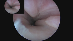
Pre bulking
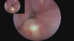
Post bulking
- Urethral Bulking for Recurrent Urinary Tract Infections in a Female Dog with Suspected Subclinical Incontinence by Tanner Slead, DVM, DACVIM - May 29, 2025
- Gastrointestinal Foreign Body by Jeremy Fleming, DVM, DACVS-SA - March 24, 2025
- Angular Limb Deformity by Russell Kalis, DVM, DACVS-SA - November 18, 2024