
Background
The most common oral tumors of dogs, in descending order of frequency, are malignant melanoma, squamous cell carcinoma, fibrosarcoma, osteosarcoma, and acanthomatous ameloblastoma. Each of these tumors has significant potential to invade surrounding tissues, including bone. These tumors vary significantly in their metastatic potential. While, malignant melanomas have a high metastatic potential, squamous cell carcinomas, and fibrosarcomas do not tend to metastasize commonly. Similar to appendicular osteosarcoma, oral osteosarcoma frequently metastasizes to the lungs, although survival times after surgical excision appear to be superior to survival times observed after amputation for appendicular osteosarcoma. Acanthomatous ameloblastoma is a nonmetastatic but locally invasive tumor that frequently invades the underlying bone and should be treated by mandibulectomy or maxillectomy.
What is needed prior to surgery?
Thoracic radiographs or computed tomography (CT) for the detection of pulmonary metastases should be obtained for any dog with a suspected oral tumor. Although the yield of this test is generally low, the presence of visible pulmonary metastases indicates an extremely poor prognosis and surgery should only be considered for palliative care.
Thorough preoperative staging of oral malignancies should include an assessment of regional lymph nodes for metastases. The canine oral cavity drains to the mandibular, parotid, and medial retropharyngeal lymph nodes; however, of these, only the mandibular node is normally palpable. Lymph node size as determined by palpation is an unreliable indicator of metastases. Cytologic examination of material obtained by fine needle aspiration of the mandibular lymph node is likely to provide an accurate assessment of that node; however, the parotid and medial retropharyngeal nodes are not readily accessible to aspiration. Blood work is also needed to ensure the patient is overall healthy.
Computed tomography is an advanced imaging modality for assessing bony and soft tissue margins. CT is particularly valuable for oral lesions whose size or location may make complete resection difficult. These include maxillary or mandibular lesions; lesions that approach or extend beyond the midline; and lesions that may encroach upon the nasal cavity, pharynx, or orbit.
Procedure
Surgical resection is recommended treatment for all oral tumors. Surgical excision of most of oral tumor, with exception of those confined to the lip margins or tongue, involves removal of bone. These procedures are called mandibulectomy (for removal of part of the lower jaw) and maxillectomy (for removal of part of the upper jaw) are most commonly indicated for the treatment of oral tumors. Many clients have a natural aversion to considering these procedures and require a thorough description of the cosmetic and functional results of surgery before making a decision. Overall, clients seem extremely pleased with the cosmetic result following the surgery, along with the rapid adaptation by the patients in prehending and chewing food. After the hair grows back, most owners comment that the surgery site is undetectable.
Various types of partial mandibulectomy and maxillectomy techniques can be performed to control local tumor growth. How much bone and soft tissue will be removed depends on CT images and invasiveness of the tumor.
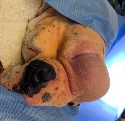
Case 1: Fig 1. Immediate pre-op caudal maxillectomy, with total orbitectomy – 5 years old neutered male Boxer – Note the large mass present on the left caudal maxilla / zygomatic arch.
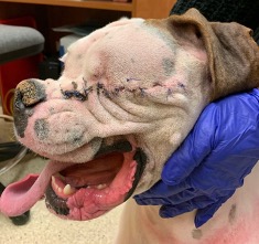
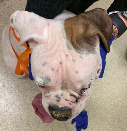
Fig 2 & 3: Two-week post-op prior to suture removal – Note that swelling is gone and asymmetry of face is minimal.
Case 2: Fig 4. Mass on the mid-caudal right mandible region on a 10 years old spayed female Great Pyrenees
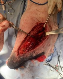
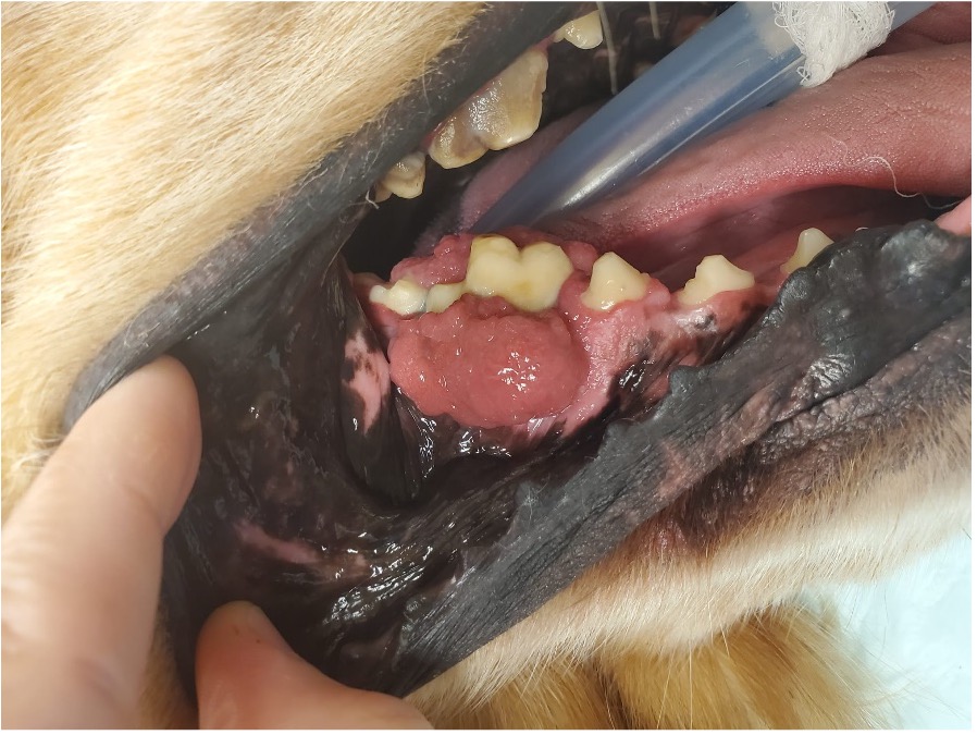
Fig 5. Photo on the left illustrates intra-op approach to the mandible.
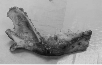
Fig. 6. Photo on the top shows the mandible after right mandibulectomy.
Fig 7. Photo showss the patient 9 months post-op.
Postoperative Care
Post-operative care after an oral surgery includes activity restriction, soft food, medications, and follow-up.
Activity Restriction – For about 2 weeks, the patient needs to have their activity restricted to not cause complications with the surgery. Too much activity can lead to bleeding, swelling, dehiscence, pain. The confinement means leash walks only, no jumping, no playing, no climbing, and no running for the majority of the recovery period. In addition, any kind of toys is prohibited since it can breakdown the sutures.
Incision Care – It usually takes about 10-14 days for the incision and soft tissues to heal. During this time, it is important to monitor the incision for any excessive swelling, oozing, or incisional dehiscence (opening up). We also recommend an E-collar (cone) placed around the dogs’ head in order to keep them from pawing at the incision. It can lead to dehiscence and/or infection of the site, which can be a serious complication.
Medications – All of patients are dispensed pain medications after surgery. The necessity for pain medication is patient dependent and can range from just a few days to the entire recovery period. In addition, antibiotics may also be prescribed. The surgeon will determine the necessary duration of pain medications or antibiotics.
Follow-up – At 2-week post-op a physical examination will be performed to ensure the activity can be resumed and e-collar removed. Further visits will depend on the histopathology of the tumor.
Potential Complications
Surgical resection is recommended treatment for all oral tumors. Intraoperative complications relate mainly to technical problems, including bleeding and anesthesia. Postoperative complications include incisional dehiscence, infection, injury to salivary ducts, subcutaneous emphysema, mandibular instability, abnormal salivation with secondary cheilitis or dermatitis, anemia, pain and discomfort, lingual dysfunction and prehension difficulties, anorexia, ocular problems, cosmetic defects, local tumor recurrence, and distant metastatic disease.
Prognosis
The prognosis for dogs undergoing these procedures has improved steadily over the years. This improvement has occurred largely because experience has shown that dogs tolerate extensive oral surgery extremely well and can experience excellent short- and long-term results if surgical candidates are chosen carefully and managed appropriately in the early postoperative period. As a result, surgeons have become more aggressive in obtaining wide resection margins. In addition, the roles of adjuvant radiation therapy in treating local disease and of chemotherapy and immunotherapy in treating metastatic disease have been better defined, and may be part of the treatment depending on histopathology results.
Jacqueline Cavalcanti, DVM, DACVS-SA
Board-Certified Small Animal Surgeon
Dr. Cavalcanti is an experienced soft tissue surgeon with emphasis in surgical oncology.
- Caustic Substance Injury by Jami Becker, DVM, DACVECC - September 30, 2024
- Management of Prolonged Starvation in a Cat –by Amaris Franco, DVM, DACVECC - June 13, 2024
- Canine Heat Stroke – by Jami Becker, DVM, DACVECC - May 20, 2024