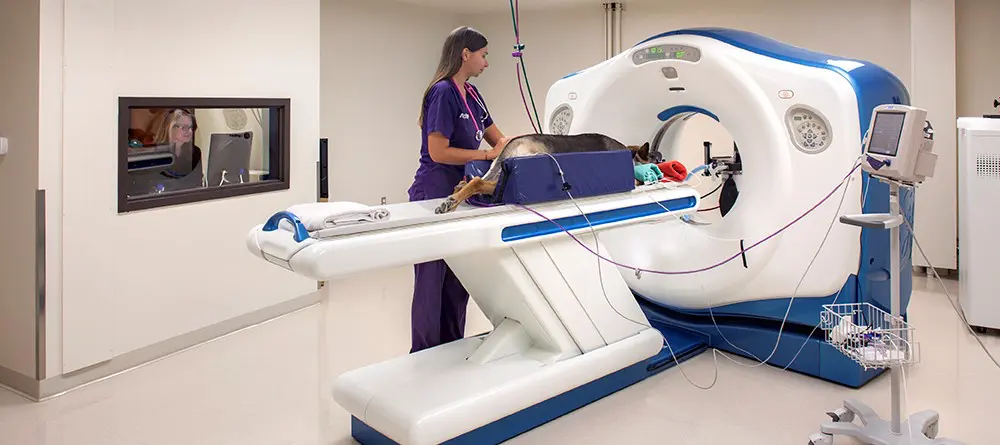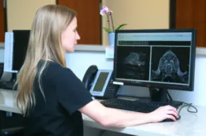
When something is amiss with your pet, we often rely on the most advanced diagnostic tools to make a diagnosis, determine the problem’s extent, and create a plan for treatment. As an integral part of this process, Austin Veterinary Emergency and Specialty Center’s advanced imaging capabilities are an invaluable part of your pet’s medical care. Advances in technology now afford your pet access to the same imaging techniques available to people at specialty medical centers, such as large universities and hospitals. In addition, we work closely with board-certified veterinary radiologists to skillfully interpret the images, for the most accurate diagnosis.
If your pet is sick or injured, and your primary care veterinarian recommends advanced imaging, we may use digital X-rays, ultrasound, computed tomography (CT), or magnetic resonance imaging (MRI) to get to the root of the problem.
Digital X-rays for pets
A digital X-ray unit produces a radiation beam that passes through your pet’s body, and is picked up by a sensor plate on the other side. The radiation is absorbed by the body tissues in differing amounts according to their density. For example, denser tissue, such as bone, absorbs much of the beam, creating a whiter area on the resulting image, whereas less-dense tissue, such as the air-filled lungs, allows most of the beam to pass through, creating darker areas.
X-rays are useful for viewing a large body area at once, and are often used as a first-line tool to pinpoint a problem area. For example, if a pet has abdominal pain, X-rays can quickly provide a concurrent view of the intestinal, urinary, and reproductive tracts. X-rays are also useful when evaluating bony abnormalities, such as fractures or arthritis, and for detecting abnormal fluid accumulations in the abdominal or thoracic cavities. Serial X-rays can be taken after contrast ingestion to observe gastrointestinal function and help diagnose foreign bodies.
Ultrasound for pets
An ultrasound probe emits high-frequency sound waves that bounce off internal body structures, are received by the unit, and translated into an image on a screen. Ultrasound produces real-time images that allow us to watch body processes, measure internal structures, and detect fluid accumulations. This modality is often used to evaluate structures in the abdomen, such as stomach wall thickness or gall bladder enlargement, and thorax, such as lung masses and abnormal lymph nodes.
By allowing us to evaluate internal structures for proper anatomy and function, ultrasound aids in the diagnosis of a variety of conditions, including:
- Internal masses, including cancer
- Pyometra
- Peritonitis
- Chronic cystitis
- Urinary stones
- Pyelonephritis
- Splenic enlargement
- Gallbladder disease
- Liver disease
Ultrasound is also commonly used to obtain tissue samples and collect urine. Using the real-time imaging, we are able to guide a needle directly to a specific site and collect samples for microscopic evaluation.
CT for pets
CT is an advanced imaging modality that gathers images of your pet’s body in thin slices, which are reconstructed into a three-dimensional image. CT scans produce images with greater detail than digital X-rays, and three-dimensional evaluation of soft tissue structures allows us to examine an abnormal organ or mass from every angle, and then plan a surgical approach. CT also provides superior images of bony abnormalities, so we can fully appreciate the extent of joint problems or bone cancer.
CT can be used to evaluate almost any body part, and aids in the diagnosis of many conditions, including:
- Masses, including cancerous tumors and metastasis
- Portosystemic shunts
- Ectopic ureters
- Inner ear disease
- Chronic nasal discharge or bleeding
- Traumatic injury
MRI for pets

An MRI unit uses a strong magnetic field and pulsed radio waves, instead of radiation, to scan a patient in thin slices to produce a three-dimensional image. MRI is the most advanced imaging technique available in human and veterinary medicine and, because a different technology is involved, can often provide information that X-rays or CT cannot. MRI is considered the gold standard for imaging nervous system structures, including the brain and spinal cord, as it provides the highest quality images, with excellent detail and resolution. MRI also provides diagnostic joint images, with superior detail of cartilage and ligaments.
Examples of conditions commonly evaluated with MRI scans include:
- Intervertebral disc disease
- Brain tumors
- Stroke
- Vascular abnormalities
With cutting-edge diagnostic tools, AVES can diagnose the most challenging medical cases, and plan for a successful recovery. Contact us to discuss advanced imaging options for your pet’s condition.
- Bacterial Meningitis – by Tracy Sutton, DVM, DACVIM - January 29, 2024
- November Oncology Continuing Education - October 10, 2023
- July Member Meeting & CE – Speaker: Jeremy Fleming - July 7, 2023
